


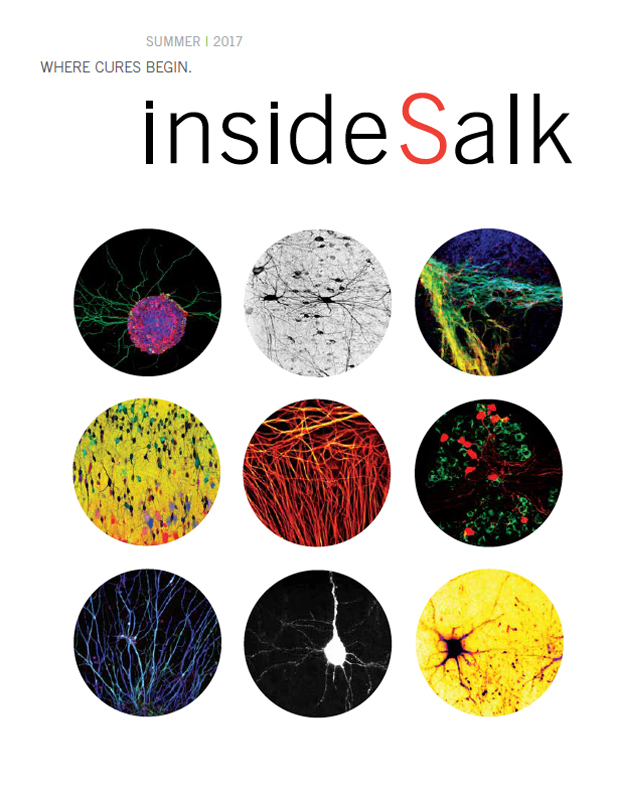


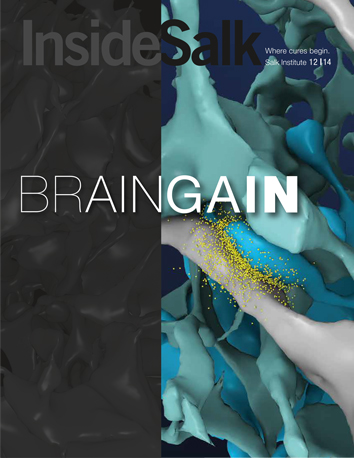


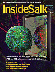


This image shows a 1.1-millimeter-thick section of mouse brain whose neurons were engineered to express tdTomato, a bright red fluorescent protein derived from Anthozoa (sea coral), allowing scientists to image all the neurons in the tissue. The sample was imaged with the Zeiss Z1 Lightsheet microscope, which boasts unprecedented speed and resolution for imaging in 3D. The Z1 was brought to the Waitt Advanced Biophotonics Center as part of a new WABC-Zeiss partnership, made possible through generous philanthropic support.
The multiple colors in this image represent different depths in 3D, with warmer colors (red) closer and cooler colors (blue) farther away from the microscope objective. Images such as these help scientists better understand the relationships between different types of brain cells, and map the connections between neurons in disparate regions of the brain.