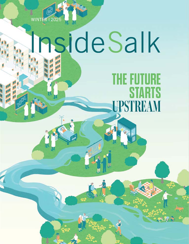
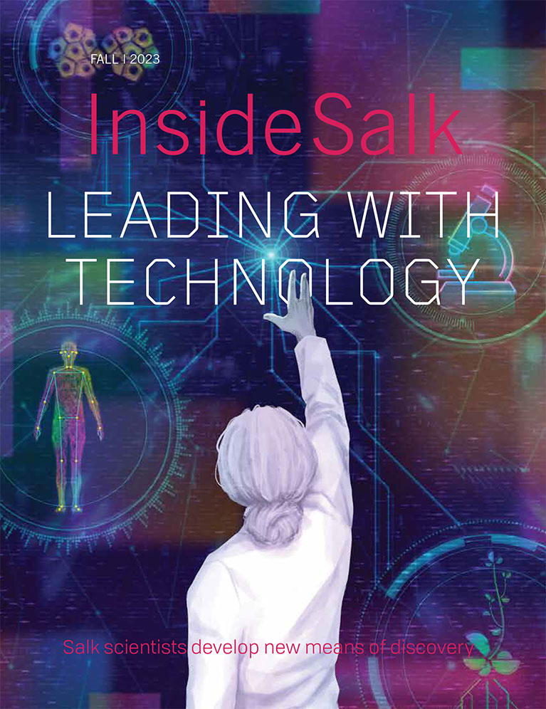


Frontiers Leading with Technology Salk scientists develop new means of discovery
[ssba]
We don’t often talk about it in these terms, but Jonas Salk’s 1955 polio vaccine—the first to be safe and effective—was a pioneering technology. Before that, only a handful of others existed, including a 1796 smallpox vaccine, an 1885 rabies vaccine, and a 1948 combined diphtheria, tetanus, and pertussis vaccine. None reached the scale of Salk’s, whose clinical trial involved more than 1.8 million children in the United States.
In developing the vaccine, Salk applied scientific knowledge to a societal problem: the annual devastation of polio, an infectious disease that primarily killed or disabled children. Thus, from the very beginning, the Salk Institute’s success has been based on its embrace of innovative technology to address humanity’s challenges.
Today, scientific research is at an inflection point driven by advances in technology and computational science. This provides new opportunities for discovery. Salk is positioned to take full advantage of the breakthroughs that computational science will enable by building the 100,000-square-foot Joan and Irwin Jacobs Science and Technology Center to house new faculty members with unique expertise, leading-edge computational and engineering technologies, and more collaborative spaces in which to do the work.
In the meantime, Salk Institute scientists are continuing to push technological limits to—among other things—store more excess atmospheric carbon in deeper plant roots, study pancreatic cancer more accurately, follow cellular activity in real time more clearly, and track all kinds of motion more easily.
The Institute’s technological advances are allowing Salk and other researchers worldwide to ask—and answer—critical questions about aging, neuropsychiatric disease, cancer, immunology, climate change, and more. In doing so, they are facilitating the future of scientific and biomedical discovery.
“I hear from people all the time who tell me how important this work is to them, for their children’s and grandchildren’s sake,” Chory adds. “Knowing we’re working to solve an existential problem for humanity is what keeps us motivated, because we’re trying to optimize for several different traits at the same time, and that’s not easy.”
PROFESSOR JOANNE CHORY
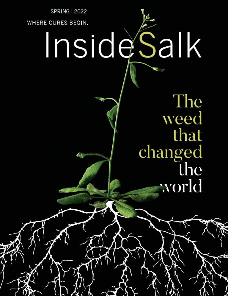
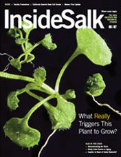
Salk Ideal Plants®: storing carbon more deeply
In the 1950s, while Jonas Salk was working in the US on the polio vaccine, across the border in Canada, plant scientists were working on a very different technology: making the seeds of the rape plant—a relative of cabbage and mustard—more edible. (The unfortunate name comes from rapa, the Latin word for turnip.) They finally succeeded in the 1970s, bringing to market the more palatable canola (the name is short for “Canadian oil, low acid”), a cultivar developed using conventional plantbreeding methods. Today, canola is the second-largest crop grown for its oil in the world, after soy.
For this reason, canola is one of the primary plants in the Institute’s Harnessing Plants Initiative, which aims to address climate change by storing excess carbon—from carbon dioxide (CO2) in the atmosphere—in deep, long-lasting plant roots. The Initiative focuses on similarly important global crops, including rice, sorghum, and soy.
“Salk Ideal Plants are the key to our Harnessing Plants Initiative’s ambitious goal of sequestering 10 gigatons of CO2 per year in plant roots,” says Professor Joanne Chory, director of the Plant Molecular and Cellular Biology Laboratory and founding director of the Initiative. Chory is also a Howard Hughes Medical Institute Investigator and Howard H. and Maryam R. Newman Chair in Plant Biology. “We’re pretty excited about this technology, which takes its cues from nature.”
A gigaton is a billion tons, and 10 gigatons is the amount of CO2 that needs to be drawn down annually to stay within 1.5°C (2.7°F) of warming by 2050, according to the National Academies of Sciences, Engineering, and Medicine.
Plant roots naturally produce and store durable, carbon-rich compounds, such as a substance called suberin; the Harnessing Plants Initiative’s goal is to dial up natural production using genetic and epigenetic methods.
Professor Wolfgang Busch, the executive director of the Initiative, is an authority on the genetics of root growth.
“Roots have the capacity to store large amounts of suberin relative to the plant as a whole, and if we put suberin and other root materials deeper in the soil, they will stay there for a long time,” says Busch, holder of the Hess Chair in Plant Science. “That’s why we are focused on finding ways to help plants make larger, deeper roots that store more suberin—these three traits are ideal for plant-based carbon sequestration.”
The objective is for the seeds of these engineered plants—trademarked as Salk Ideal Plants—to be planted at scale all over the world, serving the dual purpose of locking away carbon as the plants grow, while also producing food for Earth’s growing population.
“The natural loss of carbon from soil reduces soil quality,” says Busch. “Climate change is actually speeding up this process, which is why strategies to add carbon back into soil and keep it there longer is essential to maintain—and even increase—crop yields.”
The Harnessing Plants Initiative’s leadership team also includes Professor Joseph Ecker, Associate Professor Julie Law, Research Professor Todd Michael, and Professor Joseph Noel. The team expects Salk Ideal Plants to enrich soils with their higher carbon content and be more resilient to climate extremes, such as drought and flooding, due to the insulating quality of organic carbon.
“We are now in the double digits in terms of identifying candidate genes for each ideal trait,” says Chory. “Some of these genes are already being translated into crop plants—like canola—to be tested in field trials, and the work is only accelerating thanks to our many committed supporters.”
Multimillion-dollar donations to the Harnessing Plants Initiative have come from the Bezos Earth Fund, the Hess and Sempra corporations, and the Audacious Project (housed at TED). But individual donors have also contributed.
“I hear from people all the time who tell me how important this work is to them, for their children’s and grandchildren’s sake,” Chory adds. “Knowing we’re working to solve an existential problem for humanity is what keeps us motivated, because we’re trying to optimize for several different traits at the same time, and that’s not easy.”
Wearable microscopes: tracking cellular activity in real time during behavior
Associate Professor Axel Nimmerjahn, director of Salk’s Waitt Advanced Biophotonics Center, is well-versed in the challenges of simultaneously optimizing multiple parameters. His lab develops minuscule wearable microscopes to visualize and image the cells of the central nervous system—the brain and spinal cord—to better understand their roles in health and disease.
The spinal cord is the epicenter of critical reflexes, such as pulling our hands away from a hot stove before our brains have even registered the danger. But it is especially challenging to study the spinal cord because it is so small and difficult to access, especially in mice.
Pain and itch are sensed in the spinal cord first. When pathological, such sensations are difficult to treat, partly because the cellular mechanisms are not always clear. Since arriving at Salk in 2011, Nimmerjahn has been working to change that by making it literally easier to examine the cells involved in freely moving mice.
In 2016, with the wearable microscope his lab had developed to that point, Nimmerjahn made the surprising discovery that neurons are not the only spinal cells involved in sensing pain; star-shaped glial cells called astrocytes—long thought to be merely passive support cells for neurons—also respond to pain signals. That finding was so intriguing, the team wanted to study astrocyte activity more closely, but it was impossible with the existing technology, which was geared more toward neurons. Improving a microscope to make such studies possible was no small feat.
“The challenge on the engineering side of things is to have a microscope with cellular resolution and a large field of view,” says Nimmerjahn. “And an additional challenge is that it also needs to be small and light enough to be carried by mice—all the optical, electronic, and mechanical components have to fit into a device the size of a pinky finger that weighs no more than a few grams.”
What makes it so tricky is that different parameters can negatively influence each other—so, for example, improving a microscope to resolve small cellular structures compromises how large an area you can view. Or optimizing for both area and cellular resolution increases microscope dimensions, which becomes incompatible with experiments in freely moving animals. Or, if you manage to optimize for size and weight to enable free motion in addition to the other parameters, you may not be able to visualize cells’ activity with sufficient contrast or in multiple colors. The number of parameters that need to be optimized simultaneously to effectively image cells across large tissue areas within moving animals is daunting.
Nonetheless, in a pair of papers published this past spring, Nimmerjahn’s team reported that their latest wearable microscopes solved these complex technical problems with a highly engineered set of lenses, each just a few millimeters in diameter, whose complex combination of material properties and arrangement collectively optimized the various parameters.
“Basically, you have several tiny pieces of glass—the individual lenses,” Nimmerjahn explains. “You need to decide on the number of lenses, their thickness, diameter, curvatures, and distances between them. You need to think about what type of glass you’re using—we used different types of glass for the various lenses. Each of these glass types has a different refractive index. For example, eight different lenses, as we used in one of the microscopes, mean eight different thicknesses, diameters, glass types, and spacings, and 16 surface curvatures. So when you do modeling, you’re modeling in a very high-dimensional space.”
Modeling refers to how software creates a model of the lens system, where the researchers can vary parameters like curvature and lens separation in an effort to optimize the performance of the optical system as a whole. Different overall design variations are tried and optimized until the design achieves the requisite performance. Then the lens system and its housing can be manufactured, which is also complex since everything is so tiny. At that point, it is finally possible to maneuver the microscope over the mouse’s spinal cord and begin imaging to then optimize the tissue preparation and behavioral test parameters.
The wearable microscopes allowed Nimmerjahn’s team to visualize cellular activity in real time in the spinal cords of mice as they received different sensory stimuli. For example, the team found that astrocytes sent coordinated signals across several spinal segments when the researchers briefly but intensely squeezed the animals’ tails. Prior to the development of the new microscopes, it would have been impossible to see, in detail, what happens in cells across such broad areas of the spinal cord, much less in non-neuronal cells. Meanwhile, the team found that astrocytes show increased activity in inflammatory and chronic pain conditions, and dampening the cells’ activity alleviates pain. This makes astrocytes promising new targets for therapeutic interventions.
Nimmerjahn’s team also studies other non-neuronal cells, such as microglia (central nervous system immune cells), which play crucial roles in various central nervous system injuries and diseases, such as spinal cord injury, viral and bacterial infections, stroke, autism spectrum disorder, Alzheimer’s disease, and multiple sclerosis (MS).
MS is an inflammatory autoimmune disease affecting the brain and spinal cord, for which imaging of both neurons and non-neuronal cells could be highly revealing. In MS, a person’s immune system attacks neurons’ insulation (myelin), making it difficult or even impossible for nerve impulses to travel effectively. This can lead to myriad sensory, motor, and cognitive issues—numbness, difficulty walking, and forgetfulness, to name a few.
Nimmerjahn is one of four Salk faculty members who received $1.5 million in January from the Sol Goldman Charitable Trust to research treatments for MS. He will investigate whether it is possible to control the devastating inflammatory responses in MS through non-neuronal interventions.
“Only when you see the cellular dynamics in action can you begin to understand what’s happening, why current treatments fail, and what new or combination treatments could halt or reverse the disease,” says Nimmerjahn.
“Our lab has a strong organoid program, which enables us to grow human pancreatic cells in three-dimensional conditions, not only from normal pancreases but from pancreatic cancer as well.”
ASSISTANT PROFESSOR DANNIELLE ENGLE
Mini-organs in a dish: Studying pancreatic cancer more accurately
For most of her adult life, Assistant Professor Dannielle Engle has been dedicated to finding better diagnostic and treatment options for pancreatic cancer, the third-leading cause of cancer deaths in the US. (She shares her personal connections to the disease in Observations)
The pancreas is an abdominal organ approximately the size and shape of a small banana, which lies sandwiched between the stomach and large intestine. It makes digestive enzymes and hormones including insulin. Because symptoms of pancreatic cancer are not readily apparent, it often goes undiagnosed until it is too late. The five-year survival rate is 12.5 percent, and due to an increasing incidence, pancreatic cancer is projected to soon become the second-leading cause of cancer-related death.
Part of the reason pancreatic tumors go unseen is that they are protected within a dense meshwork of immune and connective tissue cells. Only half of the cells in a pancreatic tumor are actually cancer cells. By the time a tumor is big enough to show up on a scan, it has likely already metastasized or spread. And pancreatic tumors are hard to treat because chemotherapies have difficulty penetrating the mesh of protective cells.
Pancreatic cancer is also difficult to study. Mouse models of the disease are expensive, time-consuming to use, and not a perfect proxy for humans. Meanwhile two-dimensional experiments using human cancer cells in laboratory dishes often lack the complexity of the surrounding environment. For these reasons, Engle is devoting significant resources to a new technology for studying pancreatic cancer: pancreatic organoids—essentially, miniature human pancreases.
“Our lab has a strong organoid program, which enables us to grow human pancreatic cells in three-dimensional conditions, not only from normal pancreases but from pancreatic cancer as well,” says Engle, who holds the Helen McLoraine Developmental Chair. Her lab maintains a biobank of human pancreatic cancer and pancreatitis organoids.
This enables the team to compare healthy pancreases to diseased ones; see pancreatic cells transition from healthy to inflamed—a precursor to cancer called pancreatitis; and study the behavior of the whole collection of cells that drive pancreatic cancer development and progression.
Engle helped develop the first pancreatic organoids as a postdoctoral researcher in the lab of David Tuveson, one of the world’s foremost pancreatic cancer researchers. Since joining Salk in 2018, Engle has herself become an authority on pancreatic cancer and trains other researchers to culture “patient-derived organoids,” which are grown from cells obtained for research with the permission of their human donors.
Children with hereditary pancreatitis are a patient group close to Engle’s heart. These kids have a 50 percent lifetime risk of developing pancreatic cancer, and often have severe and painful symptoms by age 12. Engle is hopeful that patient-derived organoids will yield better therapies for such children in the next few years.
“Since we can see that these organoids maintain this inflammatory phenotype, we are asking if we can walk that back to more of a normal state using different therapeutic approaches,” says Engle. “Cancer prevention options are critical in this population. There’s one option and that’s having the entire pancreas removed. But that cuts at least 10 years off their life span, so it’s a really hard option to consider.”
Children who undergo the surgery need to have insulin-making cells transplanted back into their bodies to avoid being severely diabetic.
Engle is also working on technologies for early diagnosis of pancreatic cancer—a type of blood test, for example. In 2019, she made the consequential discovery that a carbohydrate molecule called CA19-9, which is present on many proteins, causes the progression from pancreatitis to pancreatic cancer.
She was initially hopeful that CA19-9 could be used for early detection, since CA19-9 levels are elevated in both pancreatitis and pancreatic cancer, but so far it has proven difficult. That’s because CA19-9 is not specific to those two conditions; even healthy people’s blood contains some CA19-9, and people with gallstones, liver cirrhosis, and other types of cancer can have high levels, too. The work is ongoing, buoyed by new technological advances driven by Salk and other scientists.
Since November 2022, SLEAP has been downloaded more than 75,000 times across 49 countries, resulting in more than 600 academic citations.
SLEAP: Tracking motion more easily
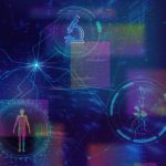
Engle’s work on a diagnostic technology for pancreatic cancer is getting an assist from Salk Fellow Talmo Pereira. Pereira is a neuroscientist by training, but at Salk he runs a cross-disciplinary computational lab. Pereira collaborates with other Salk faculty members to analyze their data using artificial-intelligence-based software he initially developed as a graduate student at Princeton University.
At Princeton, Pereira was trying to quantify the courtship dance of male fruit flies for his research. By studying animal motion, neuroscientists gain insights into such abstract mental processes as attraction, motivation, and aggression—as well as the circuitry needed for more concrete functions, like physical movement. The former can go awry in neuropsychiatric diseases such as depression and schizophrenia, while the latter can be dysfunctional in such conditions as Parkinson’s disease and obsessive-compulsive disorder.
But for Pereira, the fly quantification work was tedious and imprecise, and the options for automating it were not very good. He wondered whether the technology used by filmmakers to track motion and translate it into 3D animation—green screens and sensors—could be developed to more easily track the movements of flies. He realized that, instead of using physical sensors to map flies’ body positions, he could use computer vision (the ability for computers to understand visual images) combined with deep learning (the artificial intelligence [AI] approach of training a computer to work like the human brain).
Pereira developed the first version for tracking just one animal at a time and called it LEAP, which stands for “LEAP estimates animal poses.” (The name epitomizes a playful concept called a recursive acronym—an acronym that refers to itself.) Once Pereira arrived at Salk in November 2021, he revised LEAP to be able to track multiple animals and renamed it SLEAP, for social LEAP.
SLEAP works like this: The user identifies body parts in a few video frames or still images—for example, head (front), thorax (middle), and abdomen (rear), in the case of flies. Then the software “learns,” based on the annotated image, how to recognize flies in other images.
The software is freely available and open source, meaning anyone can inspect or alter the code as needed.
“People have used SLEAP to track every type of animal you can think of: worms, flies, mice, fish, giraffes, whales,” Pereira says. “It really spans the gamut, owing to its flexibility and also owing to the fact that we put a lot of work into making it a really nice, easy-to-use software that doesn’t require any technical expertise.”
The power of the software comes from its ability to learn, which means that after initial training the user may not need to make many corrections to its identifications before the software becomes fairly accurate. In fact, the software proved so effective at tracking animal movement, Pereira soon realized that with very little data, researchers could track any set of points, whether animals, plants, cells, or even nonliving objects.
Pereira and Professor Thomas Albright, also a neuroscientist, used SLEAP to track how humans visiting the Los Angeles County Museum of Art (LACMA) interacted with exhibitions. The study aimed to help improve exhibition design and provide insights into how humans experience the world around them. Ten cameras covered every bit of the exhibit space and recorded visitors moving through it. SLEAP tracked everything about how the people moved, from how long they lingered facing a particular exhibition to whether they interacted with other people.
Pereira is also collaborating with Professor and Hess Chair in Plant Science Wolfgang Busch, using SLEAP to track the growth and motion of plant roots, as part of the Harnessing Plants Initiative.
The SLEAP software frees up researchers to focus on analyzing data rather than on annotating it for identification. It is rapidly gaining a reputation for usefulness, and since November 2022 it has been downloaded more than 75,000 times across 49 countries. It has yielded more than 600
academic citations.
“Our user base continues to increase at an alarming rate,” Pereira laughs.
As for the pancreatic cancer diagnostic technology, Pereira, Engle, and Professor Christian Metallo are using a 2023 Salk Collaboration Grant to see whether they can use AI to predict cancer from body language.
“We have these mouse models of pancreatic cancer and we know the disease affects their metabolism in different ways, which in turn affects their behavior, particularly their feeding behavior,” says Pereira. “And so the idea is to pull out all of these little nuances of the body language of the animals that eventually develop the disease.”
In other words, there could be statistical patterns related to having cancer that a computer might be more likely than a human to find. If successful in the mouse model, Pereira envisions AI someday being used to help diagnose cancer extremely early in humans, with doctors routinely taking short videos of patients going through a series of movements for the software to analyze.
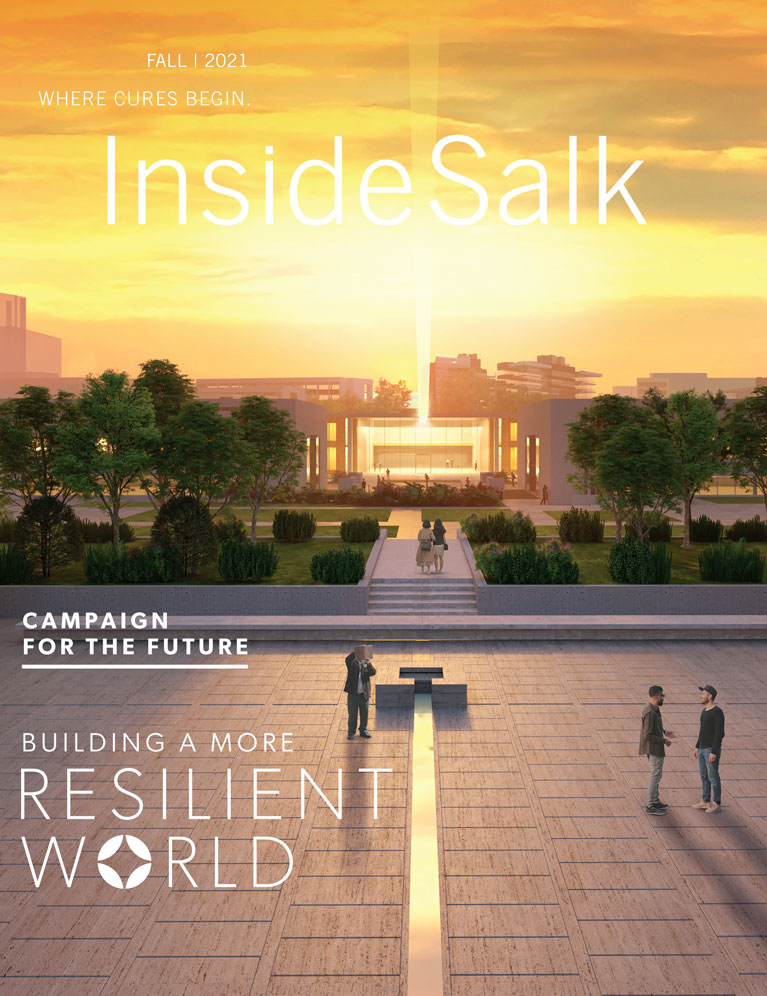
As part of the Campaign for Discovery, a seven-year, $750 million comprehensive fundraising campaign, Salk plans to build the new Joan and Irwin Jacobs Science and Technology Center on the east side of the Institute’s iconic campus. The 100,000-square-foot facility will provide much-needed research and support space to advance and expand the Institute’s scientific mission.
Joan and Irwin Jacobs Science and Technology Center: facilitating the future of discovery
The Jacobs Center will be home to Salk’s Cancer Center, Center for Healthy Aging, Hess Center for Plant Science, and Crick-Jacobs Center for Theoretical and Computational Biology. Their colocation and expansion will create more opportunities for life scientists to leverage leading-edge technologies and work side by side with engineers to build and customize research tools and design novel experiments.
The Jacobs Center will be a home for the kinds of cross-disciplinary collaborations that happen only at Salk—a neuroscientist tracking plant growth or an expert in metabolism using AI to predict cancer, for example. The Center will build on the power of Jonas Salk’s vision of flexible and functional design and will empower the next generation of scientists—those already at Salk and those yet to come.
Science is a collaborative pursuit, and we invite you to join us in developing new technologies and accelerating life-changing discoveries at salk.edu/campaign.
Support a legacy where cures begin.
Scientific discovery at the Salk Institute is made possible through your annual contributions. Your support will accelerate the pace of breakthroughs in understanding disease and pave the way to new drug therapies.
Get involved
Featured Stories
 Leading with Technology – Salk scientists develop new means of discoveryOur scientists continue to push technological limits to—among other things—store more excess atmospheric carbon in deeper plant roots, study pancreatic cancer more accurately, follow cellular activity in real time more clearly, and track all kinds of motion more easily.
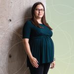 Dannielle Engle—Putting patients firstEngle, an assistant professor, has a deeply personal connection to pancreatic cancer that changed her career trajectory and made her want to focus on the disease. Inside Salk sat down with Engle to find out more about her dedication to finding better treatment options.
 Weiwei Fan—Life is energyFan, a staff scientist in Professor Ronald Evans’ lab, studies mitochondria because he feels drawn to understanding how these energy generators within our cells function and the natural variations that exist between individuals.
 Natanella Illouz-Eliaz—Recipe for a plant biologist: tomatoes, failure, and perseveranceIllouz-Eliaz, a postdoctoral researcher in Professor Joseph Ecker’s lab, grew up in Israel near the border with Lebanon, where high-pitched sirens periodically drove her family into bomb shelters for safety. But her parents insisted that she get the best education possible.
 Richard Heyman—From Salk to biotech and backHeyman, a scientist and entrepreneur who has founded numerous biotechnology companies, currently serves as vice chair of Salk’s Board of Trustees, but his Salk story actually began when he was a postdoctoral researcher in the lab of Professor Ronald Evans.