Imaging method highlights new role for cellular “skeleton” protein
While your skeleton helps your body to move, fine skeleton-like filaments within your cells likewise help cellular structures to move. Now, Staff Scientist Uri Manor and co-first authors Cara Schiavon and Tong Zhang have developed a new imaging method that lets them monitor a small subset of these filaments, called actin. They observed how actin mediates an important function: helping the cellular “power stations” known as mitochondria divide in two. The work could provide a better understanding of mitochondrial dysfunction, which has been linked to cancer, aging and neurodegenerative diseases.
Featured Stories
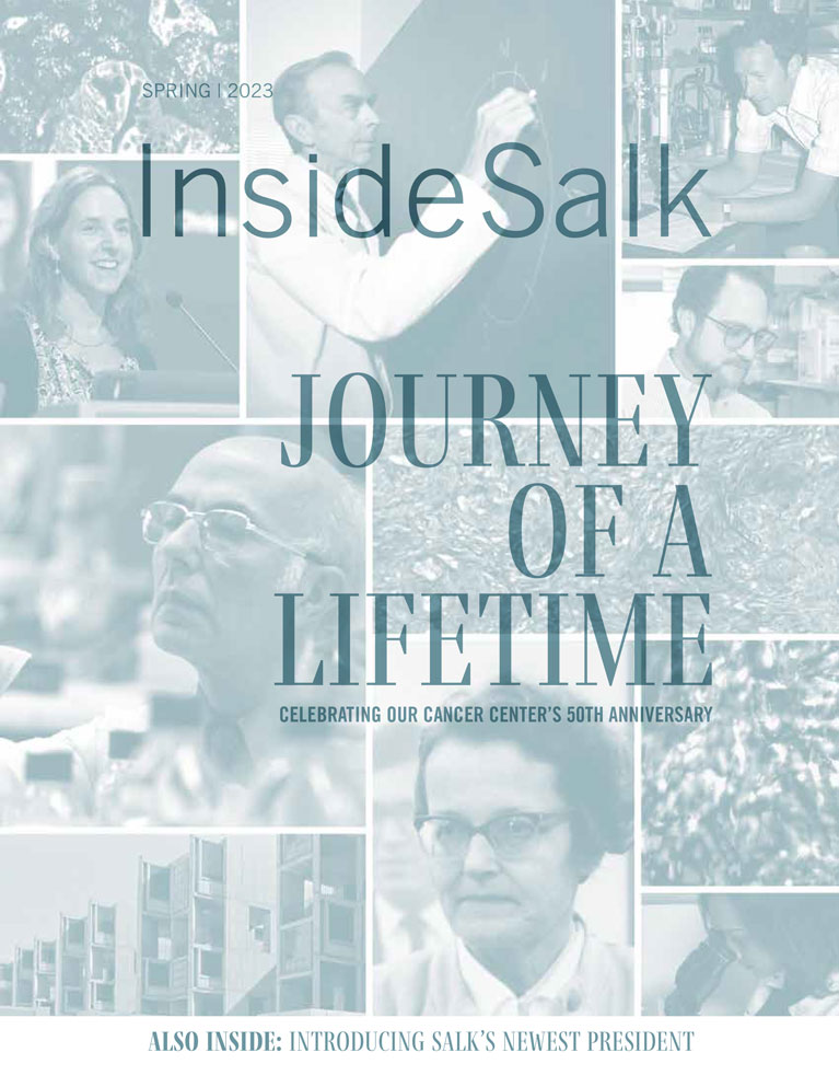
 Leading Salk science into the futureInside Salk sat down with Rusty Gage to learn more about his background, approach to managing a world-renowned Institute, and vision for Salk science over the next decade.
 Julie Law – Revealing RNAAssociate Professor Julie Law shares common themes underlying her research and discusses what motivates her both in and out of the lab.
 Gerald Pao – Pushing the limits in science and lifeFrom studying the novel coronavirus to downloading brains to computers, Staff Scientist Gerald Pao is at the forefront of scientific advancement.
 Austin ColeyAustin Coley, though only at Salk since 2019, has already taken an active role in everything from conducting innovative research on the brain to spearheading a wide variety of outreach activities.
 Salk’s Harnessing Plants Initiative (HPI) Garners Widespread SupportNew grants are supporting the Institute’s efforts to optimize plants’ natural ability to store carbon and mitigate climate change. This support bolsters the ongoing HPI project focused on model plants that was funded through donations to The Audacious Project in 2019.


